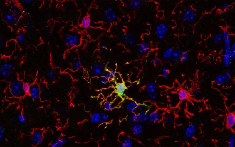
A recent study published in Nature Neuroscience indicates that, contrary to common belief, the immune cells of the brain, known as microglia, are not all the same. Researchers found that a unique microglial subset with unique features and function is important for establishing proper cognitive functions in mice. Evidence for such microglial subsets exists also for the human brain, opening exciting new possibilities for novel therapies.
An international collaboration led by researchers from University of Helsinki, Karolinska Institutet and University of Seville characterized ARG1+ microglia, a subset of microglial cells, that produces the enzyme called arginase-1 (ARG1). Using advanced imaging techniques, the team found that ARG1+ microglia are abundant during development and less prevalent in adult animals. Strikingly, these ARG1+ microglia are located in specific brain areas important for cognitive functions such as learning, thinking and memory.
“Cognition and memory are crucial components of what makes us human, and microglia are necessary for proper brain development and function. Cognitive decline is a common feature of neurodegenerative and psychiatric conditions like Alzheimer’s and Parkinson’s disease, schizophrenia and depression,” says Dr. Vassilis Stratoulias, senior researcher at the University of Helsinki and lead author of the study.
“Microglia are involved in virtually all brain pathologies, making them prime candidates for novel drug targets and innovative therapeutic approaches. Understanding the fundamental biology of these cells will be the way to produce new directions for drug development to treat currently untreatable diseases of the brain,” adds co-author, Dr. Mikko Airavaara from the University of Helsinki.
Abnormal behavior reveals cognitive deficits
The researchers found that mice lacking the microglial protein ARG1 were less willing to explore new environments. This abnormal rodent behavior is linked to cognitive deficits and, more specifically, to impairments in the hippocampus, a part of the brain important for learning and memory.
The researchers could not identify any differences in the shape of ARG1+ microglia when compared to neighboring microglia that do not express ARG1, suggesting a reason why these microglia have not been studied before. Using a technology that allows for comparison of the RNA profiles between cell populations, ARG1+ microglia were found to be significantly different from neighboring ARG1-negative microglia on the molecular level.
Another key finding of the study is that female animals exhibited more pronounced behavioral and hippocampal impairments caused by ARG1 microglial deficiency. Sex bias is present in many diseases including Alzheimer’s disease. In fact, women are more likely than men to suffer from Alzheimer’s disease—the most common neurodegenerative disease in which cognitive abilities are severely compromised.
Microglia have emerged as key players in Alzheimer’s illness in recent years, making the findings of this study relevant to this disease. Although more research is needed to demonstrate a link between Alzheimer’s disease and a specific microglial subset, this study could provide a new prism under which we see Alzheimer’s disease in specific—and brain diseases in general—and open new therapeutic pathways.
Dr. Bertrand Joseph, professor at Karolinska Institutet and senior author, says, “In addition to offering better understanding of brain development and the contribution of microglial diversity to that, the study could provide new clues about how to manage neurodevelopmental disorders or neurodegenerative disorders presenting a cognitive component and often differences between males and females.”
More information:
Vassilis Stratoulias et al, ARG1-expressing microglia show a distinct molecular signature and modulate postnatal development and function of the mouse brain, Nature Neuroscience (2023). DOI: 10.1038/s41593-023-01326-3
Journal information:
Nature Neuroscience
Source: Read Full Article
