In a recent Proceedings of the National Academy of Sciences (PNAS) study, researchers analyze T-cell reactivity following recovery from the coronavirus disease 2019 (COVID-19).

Study: Transient and durable T cell reactivity after COVID-19. Image Credit: UGREEN 3S / Shutterstock.com
Background
Naïve cluster of differentiation 4-positive (CD4+) T lymphocytes differentiate into antigen-specific effector and memory T helper (TH) cells upon antigen encounter. TH 1-polarized cells produce interferon γ (IFN-γ) and interleukin 2 (IL-2), subsequently causing cell-mediated removal of viruses and other intracellular pathogens.
In contrast, TH 2 cells produce IL-4 and IL-13, thereby facilitating the activity of B-cells and promoting post-infection tissue repair. Likewise, naïve CD8+ T lymphocytes differentiate into effector memory, central memory, and effector cells that exhibit cytotoxicity against cells infected with viruses.
About the study
The present study determined the T-cell reactivity specific to the severe acute respiratory syndrome coronavirus 2 (SARS-CoV-2). Whole-blood samples from COVID-19-recovered healthy subjects were collected one to 21 months post-recovery.
Antigen-specific TH cell cytokines were analyzed in the samples after ex vivo stimulation with peptides spanning the N-terminus of the SARS-CoV-2 spike 1 (S1) protein or complete nucleocapsid protein.
Study findings
The samples were found to generate TH 2-type cytokines IL-4 and IL-13 in response to nucleocapsid peptides until 70 days after COVID-19 recovery. These responses were not detected in non-stimulated blood samples nor in non-infected control samples stimulated by the peptides. This initial TH 2 response was concurrent with a TH 17-type response.
Notably, the levels of nucleocapsid-induced TH 2- and TH 17 cytokines were highly correlated. The results were similar when S1 peptides stimulated whole-blood samples.
Moreover, IL-12 induction by S1 and nucleocapsid peptides was contemporaneous with IL-4, IL-13, and IL-17 levels in the early months post-infection. The striking correlation between the levels of these cytokines in the early phase of infection suggests that they were produced by the same T-cells.
Follicular helper T (TFH) cells, which transcribe numerous cytokine genes, were also analyzed to explore a unified T-cell phenotype that was responsible for producing these cytokines. However, the induction of IL-21, which is the signature cytokine of TFH cells, upon stimulation by either peptide pool was weak. This finding implies that IL-21+ TFH cells might not have been the source of the multipolar cytokine response.
S1- and nucleocapsid-induced IFN-γ and IL-2 were induced in the early months after infection and showed poor or no correlation with IL-4, IL-12, IL13, and IL-17 levels. However, these cytokines persisted without declining over time and correlated well. Thus, the SARS-CoV-2-specific T-cell responses during the early and late convalescence phases are derived from different/separate T-cell clones.
Conclusions
The durability of IFN-γ and IL-2 responses reported in the current study indicate a sustained T-cell memory response after infection with SARS-CoV-2. This was congruent with recent reports that revealed the SARS-CoV-2-reactive CD4+ and CD8+ T-cell memory response post-infection.
These studies estimated the half-life of the T-cell memory response to be three to five months after recovery from COVID-19. Comparatively, the current study findings suggest that SARS-CoV-2-reactive IL-2 or IFN-γ-producing T-cells might prevail much longer than previously reported.
It was observed that a subgroup of specialized T cells (Th1 cells) that promote the destruction of virus-infected cells were active for at least 20 months after natural COVID-19. The infected patients also harbored several other types of T cells that reacted with SARS-CoV-2. These latter T cells disappeared from blood approximately two months after recovery from infection.

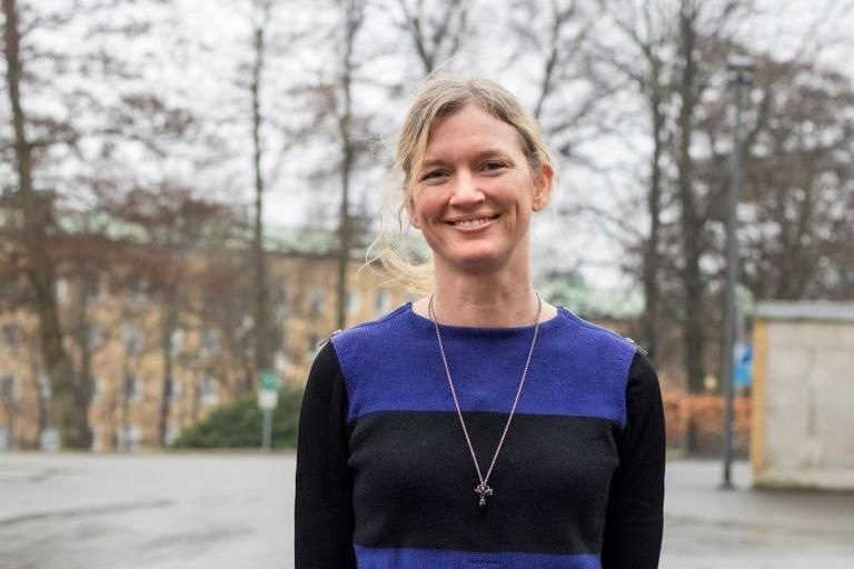
Anna Martner, Institute of Biomedicine, Sahlgrenska Academy at the University of Gothenburg. Photo: Elin Lindström
"While certain subsets of T cells disappear shortly after infection, highly specialized T cells (T helper 1 cells) remain stably present in the blood to suggest that a vital aspect of protective immunity is functional years after COVID-19", says Anna Martner, Associate Professor of immunology at the Sahlgrenska Academy. "These results may explain why re-infection with SARS-CoV-2 only rarely translates into severe COVID-19".
Some limitations of the study include its small sample size and short post-COVID-19 follow-up period. Moreover, the researchers did not explicitly identify the phenotype(s) of cytokine-generating T-cells.
- Martner, A., Wiktorin H. G., Törnell, A., et al. (2022). Transient and durable T cell reactivity after COVID-19. Proceedings of the National Academy of Sciences 119(30). doi:10.1073/pnas.2203659119
Posted in: Molecular & Structural Biology | Cell Biology | Medical Science News | Medical Research News | Disease/Infection News
Tags: Antigen, Blood, CD4, Cell, Coronavirus, Coronavirus Disease COVID-19, Cytokine, Cytokines, Cytotoxicity, Ex Vivo, Genes, Interferon, Interleukin, Intracellular, Peptides, Phenotype, Protein, Respiratory, SARS, SARS-CoV-2, Severe Acute Respiratory, Severe Acute Respiratory Syndrome, Syndrome, T-Cell

Written by
Tarun Sai Lomte
Tarun is a writer based in Hyderabad, India. He has a Master’s degree in Biotechnology from the University of Hyderabad and is enthusiastic about scientific research. He enjoys reading research papers and literature reviews and is passionate about writing.
Source: Read Full Article
