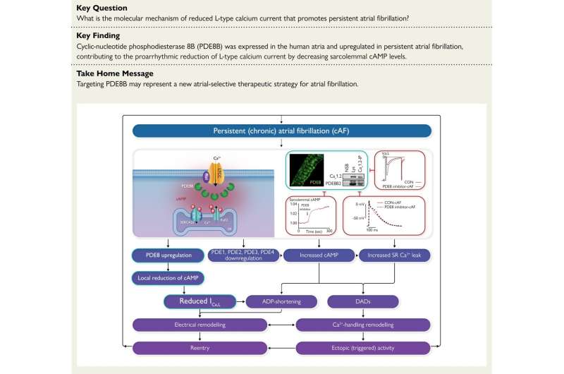
When the heart gets out of rhythm, characteristic processes occur in the heart muscle cells. Among other things, the currents of electrically charged particles (ions) change. In chronic atrial fibrillation, one of these currents is reduced. Dr. Cristina Molina from the University Medical Center Hamburg-Eppendorf (UKE) has discovered which protein is responsible for this. This provides a new and, for the first time, targeted target for drug therapies against atrial fibrillation.
Ion currents generate electrical impulses that control the heartbeat. In chronic atrial fibrillation, the influx of calcium ions into the heart muscle cells is reduced. “The reduced calcium currents are one of the characteristic features of chronic atrial fibrillation,” says Dr. Cristina Molina, a scientist at the German Centre for Cardiovascular Research (DZHK) and research group leader at the Institute for Experimental Cardiovascular Research at the UKE. “But why this is so was unclear for decades, so no therapy could be developed to address this process.”
Molina has now discovered that a protein called phosphodiesterase 8B (PDE8B) is responsible for this. In atrial fibrillation, there is too much PDE8B in the cells, more than in the muscle cells of a healthy heart. The remarkable thing is that PDE8B is only found in the cells of the atria, so these could be treated explicitly in atrial fibrillation. Conventional drugs against cardiac arrhythmias always target the whole heart, even if only the atria or only the ventricles are affected.
A knack for heart cells
There are several reasons why the cause of the reduced calcium influx in atrial fibrillation was discovered so late, although the problem has been known for a long time. One of them is that only Molina has so far succeeded in cultivating heart muscle cells from the human atrium in the laboratory over a more extended period.
Usually, these heart muscle cells die in the laboratory after a few hours. But to be able to study them adequately, they have to survive for several days. “I have been working with cells from patients’ heart muscle tissue since 2006,” says Molina. “It’s difficult because the cells differ greatly depending on the patient’s age, disease, and medication. You have to be able to deal with that.”
She and her colleagues discovered that PDE8B occurs in the human heart’s atrium a few years ago. Phosphodiesterases such as PDE8B cleave important secondary messengers in cells, leading to the removal of phosphate groups from other molecules. Since there is too much PDE8B in the atria of patients with atrial fibrillation, too many phosphate groups are removed from so-called calcium ion channels. Electrically charged particles enter the heart muscle cells through these ion channels.
Inhibitor normalizes calcium currents
There is an active substance that inhibits PDE8B and is currently being tested in a clinical trial on dementia. Molina’s research team has tested this inhibitor in the laboratory on heart muscle cells from patients with atrial fibrillation and observed that it normalizes calcium currents.
Now the plan is to test the promising substance on horses. After all, they, too, can develop atrial fibrillation just like humans. If the heart rhythm disorder occurs, the horses can no longer be used for riding. In cooperation with the Heidelberg University Hospital (Dr. Constanze Schmidt), Molina aims to treat these often “discarded” animals to see whether the inhibitor can correct atrial fibrillation. She is also pursuing a gene therapy approach because a change in the gene for PDE81 leads to too much of it being present.
The work is published in the European Heart Journal.
More information:
Nefeli Grammatika Pavlidou et al, Phosphodiesterase 8 governs cAMP/PKA-dependent reduction of L-type calcium current in human atrial fibrillation: a novel arrhythmogenic mechanism, European Heart Journal (2023). DOI: 10.1093/eurheartj/ehad086
Journal information:
European Heart Journal
Source: Read Full Article
