The superior colliculus in the mammalian brain takes on many important tasks by making sense of our environment. Any mistakes during the development of this brain region can lead to severe neurological disorders. ISTA scientist Giselle Cheung and colleagues have now, for the first time, delineated the pedigree and origin of nerve cells that make up the superior colliculus. Their findings have been published in the journal Neuron.
Cells in the brain have families, too—with all the perks and baggage that come along with them. In a human family, your ancestry influences both your body on a genetic level and your social life through your upbringing, education, and connections. For cells, their pedigree—also known as lineage—dictates their identities and how they interact and connect with each other in our developing brain.
Postdoc Giselle Cheung from the Hippenmeyer research group at the Institute of Science and Technology Austria (ISTA), together with colleagues from the Shigemoto and Siegert groups, the CeMM Research Center for Molecular Medicine, and the Medical University of Vienna, now unraveled the family relationships of neurons and glia, two major cell types in the superior colliculus.
Their findings provide insights into how disruptions in the formation of this brain region may lead to neurodevelopmental disorders like autism and attention deficit hyperactivity disorder (ADHD).
Stem cells with unrestricted potential
“The superior colliculus in the mammalian brain takes in sensory signals, especially those coming from the eyes and ears and even the sense of touch,” Cheung explains. “It processes these signals and triggers responses, for example, the unconscious movement of the eyes or the whole head. It also plays a role in maintaining focus. While being a crucial part of the brain across species, we still do not know much about its development from embryo to adult.”
Like any other organ, the brain—and the superior colliculus as part of it—develops from stem cells in an embryo. Stem cells divide and specialize further and further, eventually resulting in the enormous variety of cell types each organ needs. Now for the first time, Cheung and her colleagues shed some light on this complex process in the superior colliculus by mapping the lineage of its neural stem cells.

“We found that the development of the superior colliculus works differently than in other brain regions,” Cheung recounts the findings of the study she has been working on since she joined ISTA almost six years ago. “For example, while dividing stem cells in some brain regions take weeks to create all neurons, those in the superior colliculus quickly finish their job in just a few days.”
“While doing so, the neural stem cells in the superior colliculus retain their ability to generate any type of neuron until the end. This extraordinary capacity contrasts with stem cells in many parts of the brain, where they are specialized to only make certain groups of neurons—for example, either excitatory or inhibitory ones. In our case, we found that both kinds of neurons can be produced at the same time by the same stem cell.”
These were not the only new findings by Cheung and her colleagues. They also discovered that, during development, the layered structure of the superior colliculus is not built one layer after the other, as previously thought, but rather all at once. They also reported that a fraction of neural stem cells continue to produce glia after they have finished making neurons, a behavior similarly observed in other parts of the brain.
“All our findings together show the exceptional potential of the neural stem cells in the superior colliculus that was previously unknown to us,” adds Simon Hippenmeyer, head of the research group at ISTA. “These results help us understand how development shapes the organization and potential functions of neurons in the superior colliculus and, eventually, the entire brain. We went even further and also investigated the molecular mechanisms underpinning these developmental processes.”
Making neurons is a balancing act
After revealing what happens during the development of the superior colliculus, the researchers also tested what happens when they remove a critical gene called Pten (Phosphatase and tensin homolog) in neural stem cells. Scientists suspect that a mutated Pten gene is one of the contributing factors to autism and have shown its association with macrocephaly, the abnormal development of an enlarged brain and head.
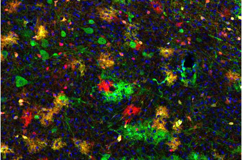
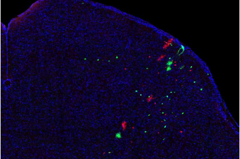
Similar to the other parts of this study, Cheung and her colleagues used a technique called Mosaic Analysis with Double Markers (MADM). MADM allowed them to mark and observe all of the progenies of individual stem cells, making them glow with distinct fluorescent colors.
Simultaneously, the Pten gene was removed in one branch of daughter cells, marked in green, but remained intact in the other, marked in red. This way, the scientists could trace and compare brain cells generated in the presence or absence of a functional Pten gene side-by-side.
The researchers found that without the Pten gene, many more inhibitory neurons were produced in the superior colliculus compared to when Pten was functional. “We showed for the first time that Pten gene function plays a role in the establishment of appropriate cell-type balance in this brain region,” Hippenmeyer adds. “We think that the disruption of such balance in the superior colliculus may lead to a deficit in the processing of sensory signals, potentially explaining disorders like autism and ADHD.”
More information:
Peter Koppensteiner et al, Multipotent Progenitors Instruct Ontogeny of the Superior Colliculus, Neuron (2023). DOI: 10.1016/j.neuron.2023.11.009. www.cell.com/neuron/fulltext/S0896-6273(23)00883-8
Journal information:
Neuron
Source: Read Full Article
