An Argonne study has revealed that short-lived mice and longer-living primates develop brain synapses on the exact same timeline, challenging assumptions about disease and aging. What does this mean for humans—and yesterday’s research?
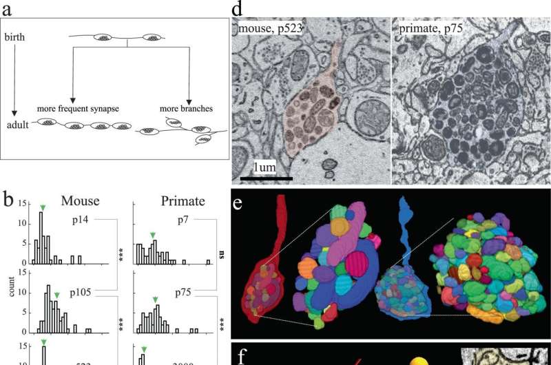
Mice typically live two years and monkeys live 25 years, but the brains of both appear to develop their synapses at the same time. This finding, the result of a recent study led by neuroscientist Bobby Kasthuri of the U.S. Department of Energy’s (DOE) Argonne National Laboratory and his colleagues at the University of Chicago, is a shock for neuroscientists. The article is published in the journal Nature Communications.
Until now, brain development was understood as happening faster in mice than in other, longer-living mammals such as primates and humans. Those studying the brain of a 2-month-old mouse, for example, assumed the brain was already finished developing because it had a shorter overall lifespan in which to develop. In contrast, the brain of a 2-month-old primate was still considered going through developmental changes. Accordingly, the 2-month-old mouse brain was not considered a good comparison model to that of a 2-month-old primate.
That assumption appears to be completely wrong, which the authors think will call into question many results using young mouse brain data as the basis for research into various human conditions, including autism and other neurodevelopmental disorders.
“A fundamental question in neuroscience, especially in mammalian brains, is how do brains grow up?” said Kasthuri. “It turns out that mammalian brains mature at the same rate, at every absolute stage. We are going to have to rethink aging and development now that we find it’s the same clock.”
Gregg Wildenberg is a staff scientist at The University of Chicago and the lead author of the study along with Kasthuri and graduate students Hanyu Li, Vandana Sampathkumar and Anastasia Sorokina. He looked closely at the neurons and synapses firing in the brains of very young mice. He marveled that the baby mouse crawled, ate and behaved just as one would expect despite having next to no measurable connections in its brain circuitry.
“I think I found one synapse along an entire neuron, and that is shocking,” said Wildenberg. “This living baby animal was existing outside of the womb six days after birth, behaving and experiencing the world without any of its brain’s neurons actually connected to each other. We have to be careful about overinterpreting our results, but it’s fascinating.”
Brain neurons are different than every other organ’s cells’ neurons because brain cells are post-mitotic, meaning they never divide. All other cells in the body—liver, stomach, heart, skin and so on—divide, get replaced and deteriorate over the course of a lifetime. This process begins at development and ultimately transitions into aging. The brain however, is the only mammalian organ that has essentially the same cells on the first and the last day of life.
Complicating matters, early embryonic cells of every species appear identical. If fish, mouse, primate and human embryos were all together in a petri dish, it would be virtually impossible to figure out which embryo would develop into what species. At some mysterious point, a developmental programming change happens within an embryo and only one specific species emerges. Scientists would like to understand the role of brain cells in brain development as well as in the physical development within species.
Kasthuri and his team were able to advance their recent discovery thanks to the Argonne Leadership Computing Facility (ALCF), a DOE Office of Science user facility. The ALCF is able to handle enormous datasets—”terabytes, terabytes and more terabytes of data,” said Kasthuri—to look at brain cells at the nanoscale. The researchers used the supercomputer facility to look at every neuron and count every synapse across multiple brain samples at multiple ages of the two species. Collecting and analyzing that level of data would have been impossible, said Kasthuri, without the ALCF.
Kasthuri knows many scientists will want more data to confirm the findings of the recent study. He himself is reconsidering past research results in the context of the new information.
“One of the previous studies we did was comparing an adult mouse brain to an adult primate brain. We thought primates are smarter than mice so every neuron should have more connections, be more flexible, have more routes and so on,” he explained. “We found that the exact opposite is true. Primate neurons have way fewer connections than mouse neurons. Now, looking back, we thought we were comparing similar species but we were not. We were comparing a 3-month-old mouse to a 5-year-old primate.”
The research’s implications for humans is blurry. For one thing, behaviorally, humans develop more slowly than other species. For example, many four-legged mammals can walk within the first hour of life whereas humans frequently take more than a year before walking first steps. Are the rules and pace of synaptic development different in human brains compared to other mammalian brains?
“We believe something remarkable, something magical, will be revealed when we are able to look at human tissues,” said Kasthuri, who suspects humans may be on a different schedule altogether. “That’s where the clock that is the same for all these other mammalian species may get broken.”
Wildenberg hopes the information gathered during the study will lead to the development of pharmaceuticals that better target human neurological disorders and disease.
“Mouse models may be great for developing cardiovascular medicines because hearts, which are basically pumps, work similarly across species,” he said. “However, developing drugs for neurological conditions is extremely hard. It’s important to understand how different species’ brains evolve so that scientists can tailor approaches based on the brain’s innovations and adaptations.”
More information:
Gregg Wildenberg et al, Isochronic development of cortical synapses in primates and mice, Nature Communications (2023). DOI: 10.1038/s41467-023-43088-3
Journal information:
Nature Communications
Source: Read Full Article
