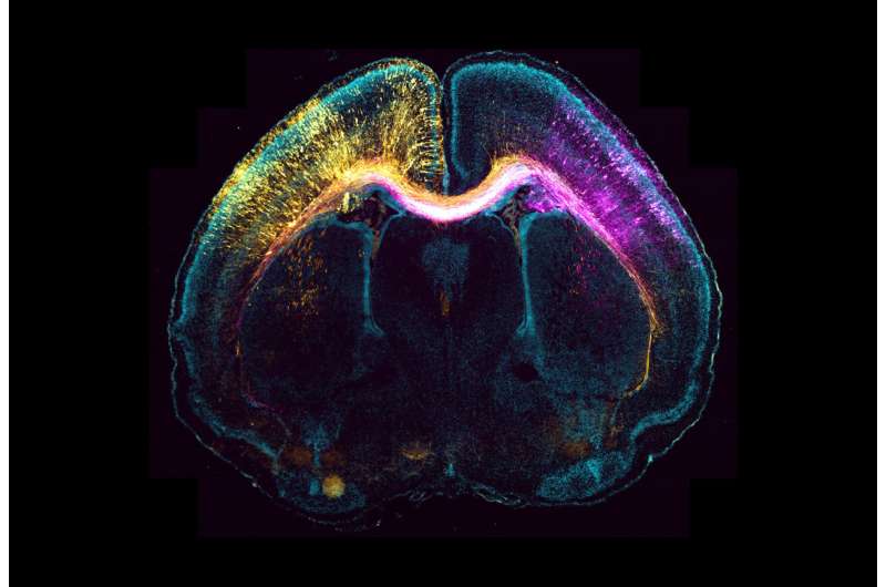
Researchers from the DZNE have solved an important puzzle in neurobiology: the wiring and the movement of nerve cells are interwoven, but separately controlled.
The study focuses on neuronal growth and migration: As nerve cells form, they wire the brain to enable communication with other nerve cells. One of these wires, the axon, becomes long; these wires are a basis for neuronal networks. At the same time, nerve cells migrate to a specific place in the brain, the cortex.
Remarkably, these dynamic processes are separately controlled: The axon continues to grow to connect with its target cells even after the nerve cell has already found its final position.
“We found that the centrosome—an organelle that drives cell division—regulates the nerve cell migration; for the formation and growth of the axon, however, it does not play a role,” Dr. Stanislav Vinopal and Dr. Sebastian Dupraz of the German Center for Neurodegenerative Diseases (DZNE) say. They are the first authors of the study, which now appears in Neuron.
Until now, experts have debated the role of the centrosome. The process of growth and migration is enabled by a dynamic skeleton of the cell, the cytoskeleton. The cytoskeleton comprises microscopic tubules, called microtubules. They form also the backbone of the axon. The microtubules can be generated by the centrosome. With their results, the participating researchers from the group of Professor Dr. Frank Bradke have solved a central puzzle in the field of neurobiology, which science has been trying to answer for years.
The fact that the growth of the axon and the control of its migratory movement are not related is an unexpected result: “Both actions occur simultaneously and both are dependent on microtubules. And still, they are controlled independently of each other,” says Stanislav Vinopal, who, after working for the DZNE, is now conducting research at Jan Evangelista Purkyne University in Usti nad Labem, Czech Republic.
For their study, the researchers developed novel molecular tools. “These molecular tools allow us to finely control the function of the centrosome to generate microtubules,” explains Sebastian Dupraz. In this way, its activity can be decreased or increased.
The scientists showed in the mouse brains that the axon form independently of the centrosomal activity. However, neuronal migration is significantly influenced. “A different mechanism is apparently responsible for the growth of the axon, the so-called acentrosomal formation of microtubules,” concludes Dupraz. “This will now become the subject of our future research.”
With their work, the scientists can now align two theories that previously contradicted each other: There were proponents of the theory that the centrosome plays a significant role in neuronal development and those who disputed it. “For our study, we disentangled the two mechanisms that occur in neurons simultaneously,” says Stanislav Vinopal. “For the growth of the axon itself, we found that the centrosome is not necessary. For the process of neuronal migration, however, it plays a major role.”
The DZNE scientists’ discovery may help develop a molecular therapy for some inherited diseases, such as so-called developmental pachygyrias, that are linked to mutations of the centrosomal protein gamma-tubulin. Also in these disease phenotypes, axons are mostly intact, while neuronal migration is impaired. “Presumably, the same molecular mechanism is behind these disorders, so a future therapy might focus on this point,” the DZNE researchers say.
More information:
Frank Bradke & colleauges, Centrosomal Microtubule Nucleation Regulates Radial Migration of Projection Neurons Independently of Polarization in the Developing Brain, Neuron (2023). DOI: 10.1016/j.neuron.2023.01.020. www.cell.com/neuron/fulltext/S0896-6273(23)00070-3
Journal information:
Neuron
Source: Read Full Article
