The brain is arguably the most crucial aspect of our existence. Our brain health governs how well we function. In turn, our brain health is determined by the blood supply to our brain via “cerebral blood flow” (CBF), which regulates the supply of oxygen and nutrients and removes metabolic by-products. An imbalance in CBF can lead to brain disorders such as headache, seizures, Alzheimer’s disease (AD), and stroke.
Observing local CBF during neural activity could, therefore, help unravel the origins of brain disorders. Speckle imaging, a technique based on the analysis of large number of short exposures, is particularly popular in this regard because it is non-invasive, label-free, simple, and provides high time resolution. However, it cannot provide information on both blood flow direction and speed, making it difficult to analyze and monitor changes in blood flow.
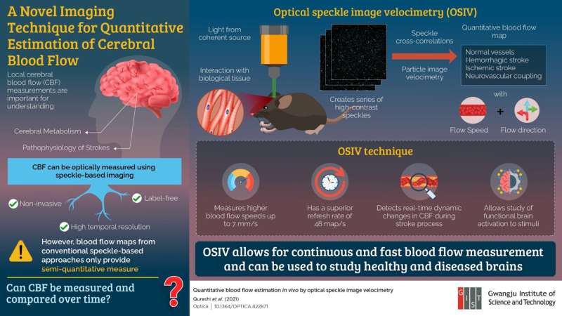
In a recent study, researchers led by Prof. Euiheon Chung from the Gwangju Institute of Science and Technology (GIST) in Korea came up with an innovative solution to this problem. The team developed a technique called “optical speckle image velocimetry” (OSIV) that creates an absolute flow map in real time with information on both speed and direction and a superior time resolution. Prof. Chung explains, “We intended to create a new technique that, unlike its predecessors, allows for a quantitative analysis of CBF and does not require complex mathematical modeling for flow measurements.” This paper was made available online on 13 August 2021 and was published in Volume 8, Issue 8 of the journal Optica.
OSIV utilizes particle image velocimetry and speckle cross-correlations to detect blood flow velocities up to 7 mm/s and can measure flow maps at up to 190 Hz. To put OSIV to the test, the team used it to image blood flow during a stroke in a mouse brain in vivo, obtaining quantitative flow measurements without needing a tracer or a high-speed camera.
Source: Read Full Article
