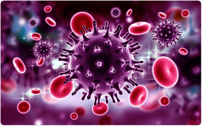
Flow cytometry is a technique commonly used to identify and separate specific cells from a mixed population. This can be applied to a large number of cells, which is ideal for further statistical analyses. Flow cytometry has been used for the detection of viruses, and this technique has been termed “flow virometry”.

Image Credit: RAJ CREATIONZS/Shutterstock.com
Furthermore, flow cytometry has been applied to sort virus particles according to infectivity, as well as to detect cells that are infected by viruses.
How did “flow virometry” develop as a technique?
Flow cytometers measure single cells by measuring how light scatters and what fluorescence is emitted when the cell is passing through a laser beam. Viruses, being significantly smaller than cells, were initially thought to be undetectable by flow cytometry.
However, by modifying a flow cytometer and using smaller volumes for observation, it became possible to detect viruses by flow cytometry. This technique was termed “flow virometry” in 2013.
The first example of a virus that was successfully detected using flow cytometry was T2 phages, which have a width of 70nm and a length of 200nm. In these early studies, the virus particles were fixed in either glutaraldehyde or formaldehyde. Subsequently, poxvirus and fixed T4 phages were also detected by flow cytometry, and the technique was further applied to detect and count viruses in marine samples.
Sample handling had to be carried out with great care and includes steps to fix and label the virus, as well as heating to improve the permeation of the dye.
Some viruses proved more difficult to detect in the initial studies, such as the ball-shaped reovirus. However, in recent years the use of flow cytometry to detect viruses has become more common, and the range of viruses which can be detected by flow cytometry has expanded to include lambda phage, herpes simplex virus, mouse hepatitis virus, human immunodeficiency virus (HIV), Nipah virus, Junin virus, vaccinia virus, dengue virus, and human cytomegalovirus.
An example of a flow virometry protocol
In a study, Brussaard, and co. used flow cytometry to detect a variety of viruses; this included HIV, herpes simplex virus, and poliovirus. These viruses were cultured with the appropriate host cells, and then the infected cells were lysed.
Some of the viruses tested, including HIV and herpes simplex virus, were partially purified from the cell lysate by centrifugation. The samples were then fixed with glutaraldehyde and then frozen using liquid nitrogen.
The fixed virus samples were then stained with various fluorescent dyes that target nucleic acids; SYBR Green I, SYBR Green II, OliGreen, and PicoGreen. The samples were then put through a flow cytometer with an argon-ion laser.
The study showed that SYBR Green I gave the highest fluorescence intensity, and viruses with a DNA genome of 48.5 to 300 kilobases could be detected by this flow virometry method. Viruses with a small RNA genome were able to be detected at the limit of detection. Further, the study showed that the method could differentiate between the different viruses.
How has technology benefited flow virometry?
The development of flow cytometer which allowed for exocytic vesicles to be detected has also benefited the field of flow virometry. One adaptation in flow cytometers which benefits the detection of viruses as well as exocytic vesicles is the increase in the power of the lasers; if the wavelength of elements in the samples are smaller than the wavelength of the light used, the samples do not refract light well. Typically, flow cytometers use lasers which are 10 to 20mW but using lasers with 300mW means that the above issue can be avoided.
One common feature of both viruses and exocytic vesicles is their small size, and this means that photons are dispersed more broadly. Most flow cytometers measure cell sizes by detecting forward light scatter, typically covering a range of 0.5˚ to 15˚. However, a significant portion of light scattered from smaller viruses or exocytic vesicles would not be detected. Adaptation in flow cytometers to allow detection of light between 15˚ and 70˚ while blocking light from 0.5˚ to 15˚ has been useful in the development of flow virometry.
Can infectivity of viruses be detected by flow cytometry?
Studies have shown that the presence of certain lipids or proteins on a viral particle, as well as their abundance, is linked to how infective the virus particle is. Gaudin and co. investigated whether flow cytometry can be used to detect differences in individual Junin virus particles.
They found that there was a correlation between the size of viral particles and the concentration of a membrane glycoprotein, and the infectivity of the virus particle. This was possible by labeling the membrane glycoprotein before analysis by flow cytometry, and this allows for the differentiation of virus particles according to their infectivity.
Can flow cytometry be used to investigate if cells have been infected by viruses?
In a study, Sivaraman, and co. investigated whether specific cell markers could be used to identify cells that had been infected by poliovirus. Here, the authors used a TAT peptide-derived molecular beacon which was delivered into infected cells, and by coupling this with flow cytometry they were able to identify cells that had been infected by poliovirus. This could potentially be developed further as a tool to diagnose viral infections quickly.
Sources
- Lippé, R. (2018) Flow Virometry: a Powerful Tool To Functionally Characterize Viruses. Journal of Virology doi: 10.1128/JVI.01765-17
- Brussaard, C. P. D. et al. (2000) Flow cytometric detection of viruses. Journal of Virological Methods https://doi.org/10.1016/S0166-0934(99)00167-6
- Gaudin, R. and Barteneva, N. S. (2015) Sorting of small infectious virus particles by flow virometry reveal distinct infectivity profiles. Nature Communications DOI: 10.1038/ncomms7022
- Sivaraman, D. et al. (2012) Use of Flow Cytometry for Rapid, Quantitative Detection of Poliovirus-Infected Cells via TAT Peptide-Delivered Molecular Beacons. Applied and Environmental Microbiology DOI: 10.1128/AEM.02429-12
Further Reading
- All Flow Cytometry Content
- Flow Cytometry Methodology, Uses, and Data Analysis
- Flow Cytometry Techniques used in Medicine and Research
- Flow Cytometry History
- Fluorescence-Activated Cell Sorting
Last Updated: Sep 30, 2020

Written by
Dr. Maho Yokoyama
Dr. Maho Yokoyama is a researcher and science writer. She was awarded her Ph.D. from the University of Bath, UK, following a thesis in the field of Microbiology, where she applied functional genomics toStaphylococcus aureus . During her doctoral studies, Maho collaborated with other academics on several papers and even published some of her own work in peer-reviewed scientific journals. She also presented her work at academic conferences around the world.
Source: Read Full Article
