What are astrocytes?
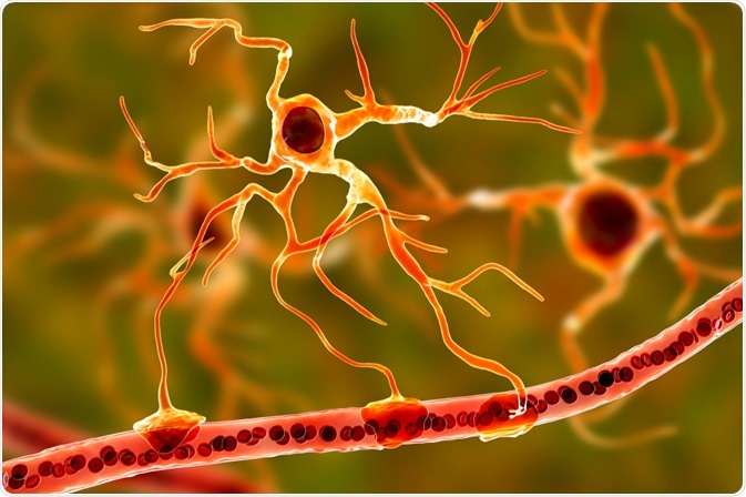
Astrocytes are highly heterogeneous neuroglial cells with distinct functional and morphological characteristics in different parts of the brain. They are responsible for maintaining a number of complex processes needed for a healthy central nervous system (CNS).
 Image Credits: Kateryna Kon / Shutterstock.com
Morphology and physiology of astrocytes
Human astrocytes are categorized based on their anatomical location and cellular morphology into protoplasmic astrocytes, fibrous astrocytes, interlaminar astrocytes, polarized astrocytes, and varicose projection astrocytes.
Protoplasmic astrocytes are found in the gray matter of the brain, while the fibrous astrocytes are found in the white matter of the brain. Interlaminar astrocytes and polarized astrocytes are present in the cortical layers nearing the white matter.
Varicose projection astrocytes also reside in the deep cortical layers. However, unlike interlaminar astrocytes, they are not found in neonatal brains.
Astrocytes form non-overlapping, tile-like sequences in the CNS and play an important role in increasing the concentration of intracellular calcium. The increase in intracellular calcium is necessary to maintain astrocyte-astrocyte and astrocyte-neuron communication and assists astrocytes in synaptic transmission.
They are involved in maintaining molecular, systemic, organ, metabolic, as well as cellular and network homeostasis. In molecular homeostasis, astrocytes regulate the release of neurotransmitter molecules such as glutamate, purines, D-serine, and Gamma Aminobutyric acid (GABA). Astrocytes also regulate the pH and K+, Ca2+, and Cl- ion homeostasis.
Astrocytes are involved in the operation of the lymphatic system, and in controlling the blood-brain barrier to maintain organ homeostasis. Metabolic homeostasis sees the involvement of astrocytes in regulating local blood flow, providing metabolic support, and glycogen synthesis and storage.
Astrocytes have a prominent role in sleep homeostasis and in the regulation of food intake and energy balance as part of systemic homeostatic support. In cellular and network homeostasis astrocytes provide support during neurogenesis, synaptic plasticity, as well as in synaptogenesis, synaptic maintenance, and elimination.
Identification of astrocytes
Glial fibrillary acidic protein (GFAP) is a prototypical marker that is used during the immunohistochemical detection of astrocytes. However, GFAP may not be present in detectable levels in many healthy CNS tissues due to the various other intra-cellular and inter-cellular signaling molecules.
Other molecular markers include S100β, glutamine synthetase, and the protein-coding gene Aldehyde Dehydrogenase 1 Family Member L1 (Aldh1L1). In addition, microarray and ribonucleic acid (RNA) sequencing techniques combined with fluorescence-activated cell sorting (FACS) or other immunopanning techniques have been used to characterize the complete mRNA profile of human astrocytes.
Fluorescent dyes such as Alexa dyes or Lucifer dyes are also used to visualize astrocytes. Probes with different spectra are also combined to enable selective visualization of astrocytes. Similarly, sulforhodamine 101 and sulforhodamine B or G are gliophilic fluorescent cationic probes that can be administered intravenously for identifying astrocytes as they penetrate the blood-brain barrier.
In recent years, a number of genetically encoded markers are being used to visualize astrocytes such as the yellow Cameleon Nano 50 (YC-Nano50).
Role of astrocytes in CNS development
Astrocytes guide the movement of axons and neuroblasts as they develop and release thrombospondin, a glycoprotein, for proper synaptic formation and functioning. Astrocytes also produce prostaglandins, arachidonic acid, and nitric oxide that help in the regulation of local blood flow in the CNS. Apart from this, astrocytes are also involved in CNS metabolism as they are known to be the main storage unit of glucose in the CNS.
Role of astrocytes in diseases
One of the hallmarks of neurodegenerative disorders is reactive astrogliosis. Reactive astrogliosis is a change observed in astrocytes at the cellular, molecular, and functional levels indicative of a CNS trauma caused either by an injury or due to the onset of a pathological condition.
Depending upon the severity of CNS damage, reactive astrogliosis has been classified into mild to moderate, severe diffuse, and severe reactive astrogliosis. In mild to moderate reactive astrogliosis, the upregulation of GFAP expression is varied along with cell hypertrophy within individual astrocytes.
In severe diffuse reactive astrogliosis, the GFAP upregulation is higher with cell hypertrophy moving beyond individual astrocytes leading to intermingling and overlapping of the processes of adjacent astrocytes. Research has demonstrated that severe reactive astrogliosis is an outcome of the brain and spinal cord injury and indicates poor prognosis, although some studies have also suggested the neuroprotective properties of scar-forming reactive astrocytes after such trauma.
Reactive astrocytes play an important role in the spread of infections, especially those of viral origin. For instance, in human herpesvirus 6 (HHV-6) encephalitis, the virus can cause encephalitis in immunocompromised as well as healthy adults. The appearance of astrocytes is of diagnostic importance especially after a stroke or in other cerebrovascular diseases such as ischemic infarct, where astrocytes are observed to surround areas of cystic encephalomalacia.
Astrocytes in therapy
The significant role that astrocytes play in neuronal health has evinced interest in researchers to look at them as novel therapeutic targets for a variety of disorders. There are many treatment methods that are being explored for targeting harmful astrocyte pathways such as nanoformulations, treatments using peptides, as well as viral gene therapy.
Approximately 20% of familial motor neuron disease is caused by the Copper-Zinc superoxide dismutase (SOD1) gene. Blackburn et al. have demonstrated that selective reduction of the gene can result in delayed onset of the disease, although its effect on the life span is less. Modulation of reactive astrocytes due to their high plasticity is one therapeutic strategy that was suggested for cell-based stroke therapy by Choudhary et al.
In addition, transplantation methodologies such as stem cell grafting to produce mature astrocytes are being investigated in mouse models for diseases such as amyotrophic lateral sclerosis (ALS). One such study transplanted human neural stem cells (NSCs) into toxin-induced Parkinson’s disease (PD) animal models and showed slowing down of the progressive symptoms of PD due to stimulation of astrocyte dedifferentiation. Another study using Huntington’s disease (HD) rats also reported differentiation of NSCs into astrocytes and neurons in the rat striatum.
With research acknowledging that astrocytes play a crucial role in neuronal health and not just a “supporting role”, many useful animal models, as well as three-dimensional matrices, are being developed for improving the therapeutic applications of astrocytes.
Sources
-
Sofroniew MV et al. (2010). Astrocytes: biology and pathology. Acta Neuropathol. doi: 10.1007/s00401-009-0619-8.
-
Verkhratsky A et al. (2017). Physiology of astroglia. Physiological Reviews. American Journal of Physiology. https://journals.physiology.org/doi/pdf/10.1152/physrev.00042.2016
-
Siracus R et al. (2019). Astrocytes: Role and Functions in Brain Pathologies. Frontiers in Pharmacology. Neuropharmacology. https://doi.org/10.3389/fphar.2019.01114
-
Blacburn D et al. (2009). Astrocyte function and role in motor neuron disease: a future therapeutic target? Glia. doi: 10.1002/glia.20848.
-
Choudhary GR et al. (2016). Reactive astrocytes and therapeutic potential in focal ischemic stroke. Neurobiol Dis. doi:10.1016/j.nbd.2015.05.003.
-
Valori CF et al. (2010). Astrocytes: Emerging Therapeutic Targets in Neurological Disorders. Trends in Molecular Medicine. https://doi.org/10.1016/j.molmed.2019.04.010
-
Tang Y et al. (2017). Current progress in the derivation and therapeutic application of neural stem cells. Cell Death and Disease. https://doi.org/10.1038/cddis.2017.504
Further Reading
- All Neurology Content
- What is Neurology?
- What is the Difference between Neurology and Neuroscience?
- What is Neuroscience?
- What is Neurosurgery?
Last Updated: Mar 9, 2020

Written by
Deepthi Sathyajith
Deepthi spent much of her early career working as a post-doctoral researcher in the field of pharmacognosy. She began her career in pharmacovigilance, where she worked on many global projects with some of the world's leading pharmaceutical companies. Deepthi is now a consultant scientific writer for a large pharmaceutical company and occasionally works with News-Medical, applying her expertise to a wide range of life sciences subjects.
Source: Read Full Article
