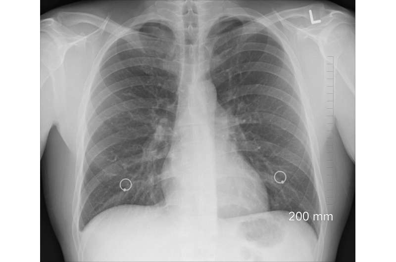
In a new publication from Cardiovascular Innovations and Applications, Zhaowei Zhu, Jianjun Tang, Xiangping Chai and colleagues from Central South University, Changsha, Hunan, China, analyze the similarities and differences of chest CT features between COVID-19 pneumonia and heart failure.
During the COVID-19 epidemic, chest computed tomography (CT) has been highly recommended for screening of patients with suspected COVID-19 because of an unclear contact history, overlapping clinical features, and overwhelmed health systems. However, there has not been a full comparison of CT for diagnosis of heart failure or COVID-19 pneumonia.
This paper describes how patients with heart failure (n = 23) or COVID-19 pneumonia (n = 23) and one patient with both diseases were assessed with clinical information and chest CT images being obtained and analyzed. No difference was found in ground-glass opacity, consolidation, crazy paving pattern, the lobes affected, and septal thickening between heart failure and COVID-19 pneumonia. However, a less rounded morphology (4 percent vs. 70 percent, P = 0.00092), more peribronchovascular thickening (70 percent vs. 35 percent, P = 0.018) and fissural thickening (43 percent vs. 4 percent, P = 0.002), and less peripheral distribution (30 percent vs. 87 percent, P = 0.00085) were found in the heart failure group than in the COVID-19 group. Notably, there were also more patients with upper pulmonary vein enlargement (61 percent vs. 4 percent, P = 0.00087), subpleural effusion (50 percent vs. 0 percent, P = 0.00058), and cardiac enlargement (61 percent vs. 4 percent, P = 0.00075) in the heart failure group than in the COVID-19 group. More fibrous lesions were also found in the COVID-19 group, although there was no statistical difference (22 percent vs. 4 percent, P = 0.080).
Source: Read Full Article
