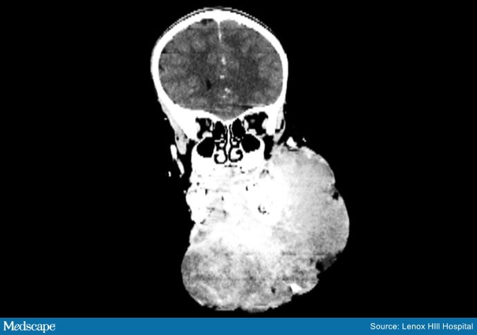
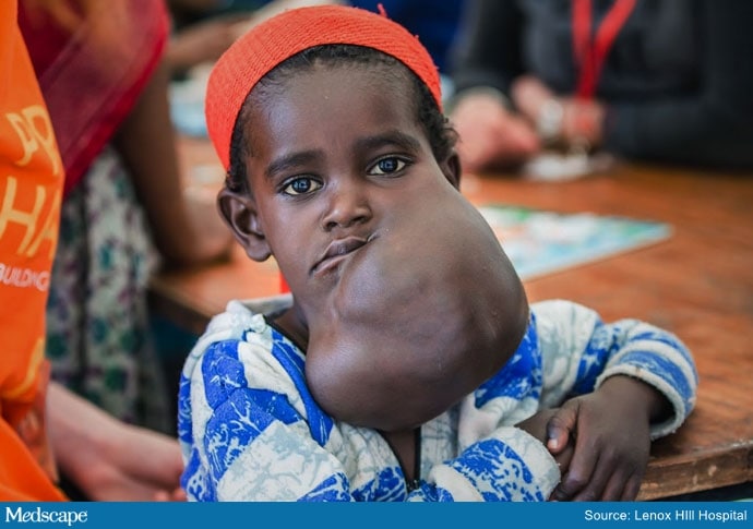
In 2019, doctors in London saw a 5-year old girl from rural Ethiopia with an enormous tumor extending from her cheek to her lower jaw. Her name was Negalem and the tumor was a vascular malformation, a life-threatening web of tangled blood vessels.
Surgery to remove it was impossible, the doctors told the foundation advocating for the girl. The child would never make it off the operating table. After a closer examination, the London group still declined to do the procedure, but told the child’s parents and advocates that if anyone was going to attempt this, they’d need to get the little girl to New York.
In New York City, on 64th street in Manhattan, is the Vascular Birthmark Institute, founded by Milton Waner, MD, who has exclusively treated hemangiomas and vascular malformations for the last 30 years. “I’m the only person in the [United] States whose practice is exclusively [treating] vascular anomalies,” Waner tells Medscape.
Waner has assembled a multidisciplinary team of experts at the institute’s offices in Lenox Hill — including his wife Teresa O, MD, a facial plastic and reconstructive surgeon and neurospecialist. “People often ask how the hell do you spend so much time with your spouse?” Waner says. “We work extremely well together. We complement each other.”
O and Waner each manage half of the cases at VBI. And in January they received an email about Negalem. After corresponding with the child’s advocate and reviewing images, they agreed to do the surgery, fully aware that they were one of only a handful of surgical teams in the world who could help her.

An image of a scan of the patient before surgery.
The challenge with vascular malformations in children, Waner says, is that they have a fraction of the blood an adult has. Where adults have an average of 5 L of blood, a child this age has only 1 L. To lose 200 or 300 mL of blood, “that’s 20 or 30% of their blood volume,” Wander said. So the removal of such a mass, which requires a meticulous dissection around many blood vessels, carries a high risk of the child bleeding out.
There were some logistical hurdles, but the patient arrived in Manhattan in mid-June, at no cost to her family. The medical visa was organized by a volunteer who also work for USAID. Healing the Children Northeast paid for her travel and the Waner Kids Foundation paid for her hotel stay. Lenox Hill Hospital and Northwell Health covered all hospital costs and post-surgery care. And Drs. O and Waner did the planning, consult visits, and procedure pro bono.
The surgery was possible because of the generosity of several organizations, but the two surgeons still had a limited time to remove the mass. Under different circumstances, and with the luxury of more time, the patient would have undergone several rounds of sclerotherapy. This procedure, done by interventional radiologists, involves injecting a toxin into the blood vessels which causes them to clot. Done prior to surgery it can help limit bleeding risk.
On June 23, the morning of the surgery, the patient underwent one round of sclerotherapy. However, it didn’t have the intended effect, Wander said, “because the lesion was just so massive.”
The team had planned several of their moves ahead of time. But this isn’t the sort of surgery you’d find in a textbook. Because it’s such a unique field, Waner and O have developed many of their own techniques along the way. This patient was much like the cases they treat every day, only “several orders of magnitudes greater,” Waner said. “On a scale of 1 to 10 she was a 12.”
The morning of the surgery, “I was very apprehensive,” Waner recalled. He vividly remembers the girl’s father repeatedly kissing her to say goodbye as she lay on the operating table, fully aware that this procedure was a life-threatening one. And from the beginning there were challenges, like getting her under anesthesia when the anatomy of her mouth, deformed by the tumor, didn’t allow the anesthesiologists to use their typical tubing. Then, once the skin was removed, it became clear how dilated and tangled the involved blood vessels were. There were many vital structures tangled in the anomaly. “The jugular vein was right there. The carotid artery was right there,” Waner said. It was extremely difficult to delineate and preserve them, he said.

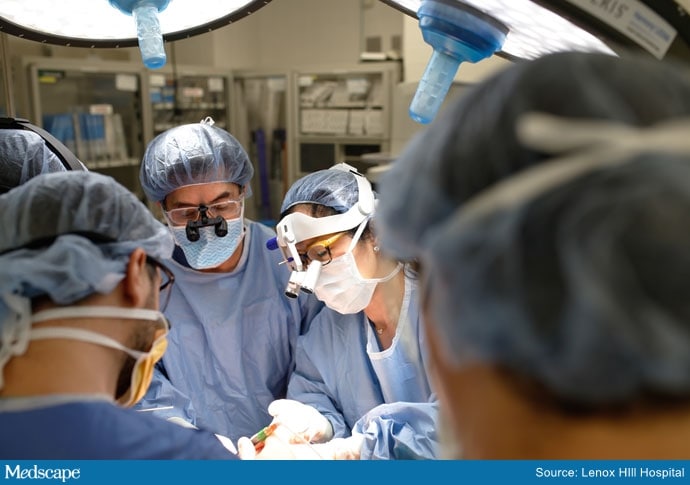
Dr Milton Waner and Dr Teresa O during the 12-hour surgery.
“That’s why we really took our time. We just went very slowly and deliberately,” O said. The blood vessels were so dilated that their only option was to move painstakingly slow — otherwise a small nick could be devastating.
But even with the slow pace the surgery was “excruciatingly difficult,” Waner said. And early on in the dissection he wasn’t quite sure they’d make it out. The sclerotherapy hadn’t done much to prevent bleeding. “At one point every millimeter or two that we advanced we got into some bleeding,” Waner said. “Brisk bleeding.”
Once they got into the surgery they also realized that the growth had adhered to the jaw bone. “There were vessels traversing into the bone which were hard to control,” O said.
But finally, both doctors realized they’d be able to remove it. With the lesion removed they began the work of reconstruction and reanimation.
The child’s jaw and cheek bone had grown beyond their normal size to support the growth. They had to shave them down to achieve facial symmetry. The tumor had also inhibited much of the child’s facial nerve control. With it gone, O began the work of finding all the facial nerve branches and assembling them to reanimate the child’s face.
Before medicine, O trained as an architect, which, according to Waner, has equipped her with very good spatial awareness — a valuable skill in the surgical reconstruction phase. After seeing a lecture by Waner, she immediately saw a fit for her unique interest and skill set. She did fellowship training with Waner in vascular anomalies, and then went on to specialize in facial nerve reanimation. The proof of O’s expertise is Negalem’s new, beautiful smile, Waner said.
The surgery drew out over 8 hours, as long as a day of surgeries for the two doctors. When O finally walked into the waiting room to inform the family of the success, the first words out of the father’s mouth were, “Is my daughter alive?”
A growth like Negalem had is not compatible with a normal life. Waner’s mantra is that every child has the right to look normal. But this case went beyond aesthetics. If the growth hadn’t been removed, the child was only expected to live 4 to 6 more years, Waner said. Without the surgery, she could have suffocated, starved without the ability to swallow, or suffered a fatal bleed.

Negalem, 5, before her surgery.
O and Waner are uniquely equipped to do this kind of work, but both are adamant that treating vascular anomalies is a multidisciplinary, multimodal approach. Specialties in anesthesiology, radiology, lasers, facial nerves — they are all critical to these procedures. And often patients with these kinds of lesions require medical and radiologic interventions in addition to surgery. In this particular case, from logistics to post-op, “it was a lot of teamwork,” O said, “a lot of international teams coming together.”
Though extremely difficult, “in the end the result was exactly what we wanted,” Waner said. Negalem can live a normal life. And as for the surgical duo, both feel very fortunate to do this work. O said, “I’m honored to have found this specialty and to be able to train with and work with Milton. I’m so happy to do what I do every day.”
Donavyn Coffey is a freelance journalist who covers health and the environment from her home in the Bluegrass. Her work has appeared in Popular Science, Insider, and SELF.
For more news, follow Medscape on Facebook, Twitter, Instagram, YouTube, and LinkedIn
Source: Read Full Article